Hip Impingement
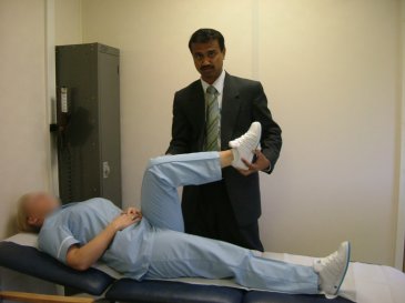
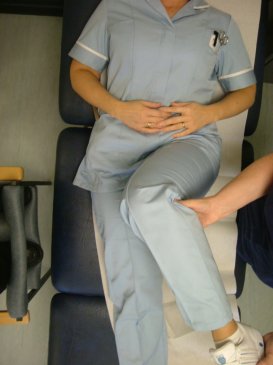
Hip Joint is a ball-and-socket joint. During normal movements of the hip, the ball glides within the socket in a smooth fashion. In instances where there is a physical abnormality in the hip joint, this movement results in an impingement at the margin of the socket. The abnormality in the hip joint can vary, but it is commonly either in the form of a bump (cam) over the ball portion of the hip joint or an abnormal over covering (pincer) of the ball by the socket. The soft tissue (Labrum) attached at the margin of the socket sustains injury due to repeated impingement. The impingement if untreated can also result in damage to the lining of the socket and thereby may initiate the wear and tear changes (arthritis) in the hip joint. During clinical examination, these patients show positive impingement test. This test is performed with patient in supine position on a couch. The affected hip is flexed to 900, adducted and internally rotated (Figure 1). Reproduction of symptoms by this manoeuvre constitutes positive impingement test.
Figure 1: Impingement Test
This clinical condition is further assessed by X-Rays (Figure 2) and MRI scans (Figure 3).
Figure 2(a) shows normal Right Hip, Figure 2 (b) shows features of bilateral Hip impingement. The contour of the head neck junction is altered.
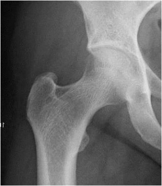
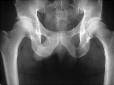
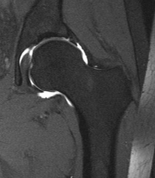
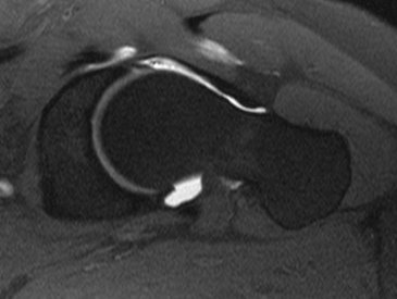
Figure 3: MRI Arthrogram show evidence of labral tear and cam impingement in the Left Hip.
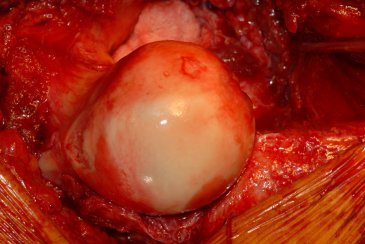
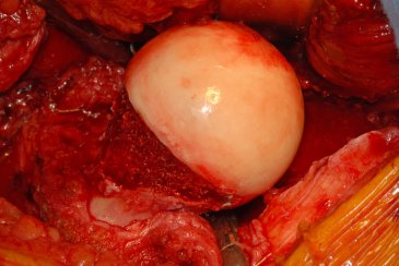
The treatment choices generally include conservative treatment with physiotherapy along with activity modification. Surgery is indicated in patients with failed conservative treatment. Surgical modalities include hip arthroscopy or open procedure to reshape the ball portion of the hip joint and address the socket issues at the same time (Figure 4).
Figure 4: Intraoperative picture showing reshaping of the ball portion of the hip joint.
The results of this specialist procedure largely depend on the pre-existing changes in the hip joint. Patients treated in the early phases of their disease do well and can return to high activity levels. A period of up to six weeks of mobilisation with the aid of crutches is advised following this procedure. The choice of treatment in hip impingement depends on the extent of associated damage to the hip joint. In the presence of significant arthritis, if the symptoms are severe, hip replacement remains the treatment of choice.
Frequently Asked Questions
What is impingement?
This is a clinical condition where the ball portion of the hip joint impinges against the socket during some of the hip joint movements.
What are the consequences of Impingement?
It is being recognised that the impingement if continues unabated may result in arthritis of the hip joint. However, there is no evidence till to-date to support that, the procedures done to relieve impingement prevent arthritis in the future.
Is it must for me to undergo surgery?
NO. The surgery is done only if you have groin pain and it is affecting your activities and quality of life. It is considered only after failed conservative therapy.
What are the types of available surgeries?
The surgery is commonly done through key hole (Hip Arthroscopy). Occasionally in some cases open surgery is used to address complex pathology. The key hole surgery is performed usually with two small cuts over your hip joint. The impingement is addressed by using instruments through these small cuts. The open surgery involves opening the hip joint and addressing the same issues under direct vision.
When can I go home after Surgery?
You will be allowed to go home either on the same day or the next day following key hole surgery. After open surgery, you will usually stay in the hospital for 2-3 days.
You are required to do partial weight bearing (20% body weight) with crutches for a period of two weeks after your key hole surgery. This will sometimes be different depending on the details of the procedures performed at the time of key hole surgery. After open surgery you are expected to be on crutches for a period of six weeks.
What are the complications of Arthroscopic (Key Hole) surgery?
The complications of hip arthroscopy are generally related to the traction employed during the procedure. These are rare indeed. These could include transient nerve paralysis in the leg, bruising and numbness in the perineal area.
What are the complications of Open surgery?
These include infection (<1%), fracture neck of femur (rare), avascular necrosis (rare). During open surgery, a piece of bone is reattached with three screws. In some patients, these screws cause local irritation and need removed in the future.
How is arthroscopy of the hip different from arthroscopy of other joints e.g., Knee?
Hip joint is a deep seated ball and socket joint. It is difficult to get an easy access. Traction applied through a boot fastened over the foot is employed to access the joint. The procedure is performed under x-ray guidance.
What is the reported success rate after these procedures?
The reported success rates range from 70% onwards. The outcome largely depends upon the degree of wear and tear changes in the hip joint.
Do I need Hip replacement in the later life?
The only indication for hip replacement will be presence of significant wear and tear (arthritis) changes accompanied by significant symptoms. Hip arthroscopy is avoided in patients who have presence of moderate to severe degenerative changes in the hip joint.
 About the Glasgow Hip Clinic